Couples who have PGD will undergo an in vitro fertilization (IVF) cycle to create embryos. Genetic analysis will then be performed on cells from each embryo prior to transfer into the woman’s uterus. To analyze an embryo, we biopsy the embryo around the third day of its development when the embryo has approximately 6-8 cells. One or two cells are taken from the embryo. The embryo is incubated until testing is complete.
The biopsied cells are analyzed using a technique called fluorescence in-situ hybridization or FISH. This technique uses probes, small pieces of DNA that are a match for the chromosomes we want to analyze, to count the chromosomes present.
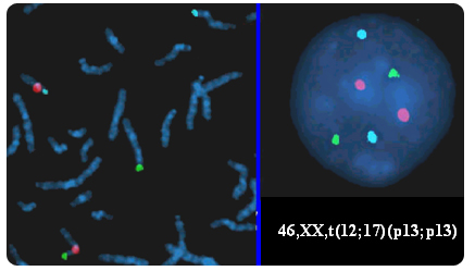
The biopsied blastomeres are first fixed to a microscope slide and then the cellular material is digested away leaving the nucleus, which contains the DNA, in a spread out and decondensed form. These cells are then hybridised with DNA probes, labeled with different fluorochromes. Each of these probes are specific for part of a chromosome; they will only attach to their exact DNA match on a particular chromosome. Excess probe is washed off, and the cell is examined under the fluorescent microscope. We then count the number of chromosomes of each type (color) there are in that cell. The geneticist therefore can distinguish normal cells from cells with aneuploidy.
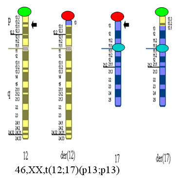
For couples undergoing IVF and PGD for translocations, the embryos will first be tested for unbalanced translocations. If technically possible, we will also perform a second test for chromosomes 13, 18, 21, X, and Y which are those chromosomes most likely to result in a liveborn child with a chromosome abnormality. The FISH analysis involves two rounds of testing for each cell.
Testing of the cells destroys them because they must be glued to a glass slide and repeatedly heated and cooled. As such, one cannot use them for another purpose or return them to the embryo. The slides are kept for future reference. This analysis causes no extra inconvenience to the patient as it is accomplished in one day.
The FISH technique is considered to have an error rate between 5 and 10%. The main problem of the use of FISH to study the chromosomal constitution of embryos is the elevated mosaicism rate observed at the human preimplantation stage. Sandalinas and collaborators found that up to 70% of the embryos they studied by FISH were mosaic for some kind of chromosomal abnormality (Sandalinas et al., 2001).